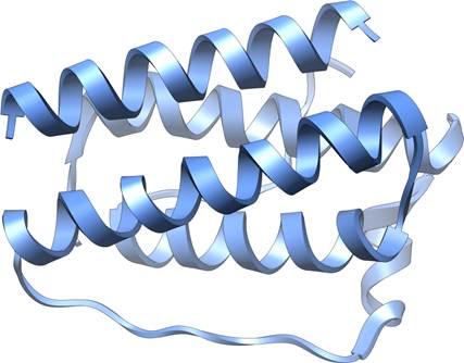
• Leptin Structure

    Leptin structure

    In the mid-1990s, a breakthrough in obesity research fundamentally changed our understanding of how the body regulates weight, hunger, and metabolism. The discovery of leptin, a hormone that would prove central to energy balance, emerged from decades of research involving an unusual laboratory mouse strain and culminated in findings that would help patients with rare but devastating metabolic disorders. This is the story of how scientific curiosity, persistence, and a colony of obese mice led to one of the most significant discoveries in modern endocrinology.

    The Mystery of the OB Mouse

    OB Mouse is a spontaneously occuring mutation in mice that caused obese.  The gene involved was the Leptin gene

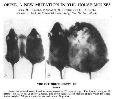
    The story begins in 1949 at the Jackson Laboratory in Bar Harbor, Maine, where researchers noticed something extraordinary: a spontaneous mutation had produced mice that became massively obese. These mice, which came to be known as the “ob/ob” mice (short for “obese”), weighed nearly three times as much as their normal littermates. They were voraciously hungry, ate constantly, and developed severe obesity along with diabetes-like symptoms.

    For decades, these ob/ob mice remained a biological curiosity. Scientists knew the obesity was caused by a recessive genetic mutation, meaning both copies of a particular gene had to be defective for the condition to appear, but the identity and function of that gene remained elusive. The mice became an invaluable research tool, but the fundamental question persisted: what was different about these animals at the molecular level?

    Adding to the intrigue was the existence of another mutant mouse strain called “db/db” (for “diabetes”), discovered in the 1960s. These mice had a remarkably similar phenotype to the ob/ob mice—they too were obese, constantly hungry, and diabetic. Yet they had a different genetic mutation. The relationship between these two strains would prove crucial to understanding the biology of weight regulation.

    Parabiosis: A Clue from Connected Mice

    Parabiosis.  What would happen if you physically connect the circulation of an OB mouse with an Normal ,mouse.  Would you correct the defect in OB mouse?  How would the normal mouse respond?

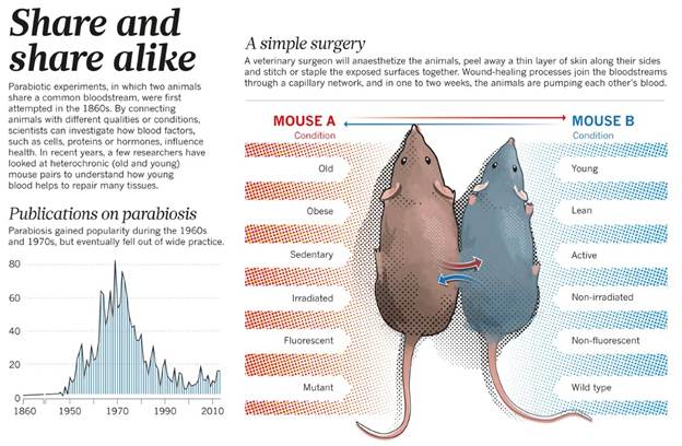
    A pivotal insight came from elegant experiments conducted in the 1970s by Douglas Coleman at the Jackson Laboratory. Coleman performed parabiosis experiments—surgically joining two mice so they shared a circulatory system. When he connected an ob/ob mouse to a normal mouse, something remarkable happened: the obese mouse ate less and lost weight. However, when he connected a db/db mouse to a normal mouse, the normal mouse stopped eating and became emaciated, while the db/db mouse remained obese.

    These results suggested a powerful hypothesis: the ob/ob mice lacked some circulating factor that suppressed appetite and promoted energy expenditure, while the db/db mice produced this factor but couldn’t respond to it. In other words, ob mice couldn’t make the signal, while db mice couldn’t receive it. The circulating factor was being produced by the normal mouse and affecting the ob mouse through their shared bloodstream, but the db mouse appeared to be overproducing the factor to no effect, overwhelming its normal partner.

    Coleman’s work provided a conceptual framework, but identifying the actual molecule would require tools that didn’t yet exist. The answer would have to wait for the molecular biology revolution.

    Jeffrey Friedman and the Hunt for the OB Gene

    Enter Dr. Jeffrey Friedman, a physician-scientist at Rockefeller University who became fascinated with understanding the genetic basis of obesity. In the late 1980s, Friedman set out to identify and clone the ob gene using an approach called positional cloning, a painstaking technique that involves tracking down a gene based solely on its chromosomal location without any prior knowledge of what the gene does or what protein it produces.

    This was an audacious undertaking. The mouse genome had not been sequenced. The techniques for positional cloning were still being developed. The project would require mapping the mutation to a specific chromosomal region, narrowing down that region through genetic linkage analysis, identifying candidate genes, and then proving which one was responsible for the obesity phenotype.

    Friedman’s laboratory spent years on this pursuit. Using genetic markers and analyzing thousands of mice from breeding crosses between ob/ob mice and normal mice, they progressively narrowed down the location of the ob gene on mouse chromosome 6. They eventually identified a region containing only a handful of genes. The team then systematically examined each candidate gene, looking for differences between normal mice and ob/ob mice.

    The breakthrough came in 1994. After nearly eight years of intensive research, Friedman’s team identified the ob gene. The gene coded for a previously unknown protein that was expressed primarily in white adipose tissue—body fat. In ob/ob mice, the gene contained a mutation that prevented production of functional protein. The team named the protein “leptin,” derived from the Greek word “leptos,” meaning thin.

    The Science Behind the Discovery

    The identification of leptin represented a triumph of molecular biology techniques. Friedman’s team used several key approaches:

    Genetic Mapping: By breeding ob/ob mice with normal mice from different strains, researchers could track which genetic markers co-segregated with the obesity phenotype across generations. This allowed them to progressively narrow the chromosomal region containing the ob gene from millions of base pairs down to a manageable segment.

    Physical Mapping: Once the region was identified, the team created overlapping clones of DNA fragments covering that area—a technique called “chromosome walking.” This allowed them to systematically examine the DNA sequence of the candidate region.

    Candidate Gene Analysis: Within the narrowed region, researchers looked for sequences that resembled known genes. They identified several candidates and examined their expression patterns and sequence variations between normal and obese mice.

    Mutation Analysis: The crucial evidence came when they found that ob/ob mice had a mutation in one particular gene that caused a premature stop signal in the protein-coding sequence. This meant the gene couldn’t produce a functional protein. A different natural mutation in another strain of obese mice affected the same gene differently but with the same result—no functional leptin.

    Expression Studies: The researchers demonstrated that the ob gene was expressed primarily in adipocytes (fat cells) and that the level of expression correlated with the amount of body fat. They showed that normal mice produced leptin in proportion to their fat stores, while ob/ob mice produced none.

    The final proof came from administering leptin to ob/ob mice. When given injections of the leptin protein, these previously insatiable, obese mice ate less, became more active, and lost weight dramatically. Their metabolism normalized. It was a stunning validation of decades of hypothesis and years of painstaking molecular work.

    What Does Leptin Do?

    The discovery of leptin revealed a sophisticated system for regulating body weight and energy balance. Leptin functions as an adiposity signal, essentially, fat tissue uses leptin to communicate with the brain about the body’s energy stores.

    Leptin is present in Adipose cells.  More Leptin means more fat in fat cells.  Leptin  enters the blood stream and binds its receptor in the hypothalamus causing metabolic changes, includeing decreasing appetite and food intake and burning fat (thermogenesis).

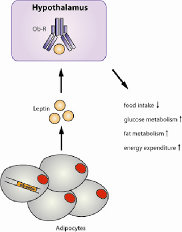
    Here’s how the system works: adipocytes produce leptin in proportion to the amount of fat they contain. More body fat means more leptin production. The leptin circulates in the bloodstream and travels to the brain, where it binds to receptors concentrated in the hypothalamus, a region critical for regulating appetite, metabolism, and energy expenditure.

    When leptin levels are adequate, the brain receives the signal that energy stores are sufficient. This triggers several responses: appetite decreases, energy expenditure increases through mechanisms like thermogenesis (heat production), and the body shifts toward using stored energy. The hypothalamus also regulates other hormones and physiological processes to maintain energy balance.

    Conversely, when leptin levels drop—as happens during weight loss or starvation—the brain interprets this as a signal that energy stores are depleted. Hunger increases, metabolism slows to conserve energy, and the body shifts toward storing any available calories as fat. This is why losing weight becomes progressively harder—the body fights to defend its fat stores through leptin-mediated mechanisms.

    The db/db mice, it turned out, had mutations in the leptin receptor gene. They produced leptin, often at very high levels, but their brains couldn’t respond to it. This explained Coleman’s parabiosis results: the circulating leptin from db mice entered the normal mice and suppressed their appetite, while the db mice themselves remained obese because they couldn’t sense the hormone.

    From Mice to Humans: The Leptin Connection

    Friedman’s discovery immediately raised a critical question: was leptin relevant to human obesity? The answer proved complex. In 1997, researchers identified the first humans with congenital leptin deficiency, children from consanguineous families who, like ob/ob mice, had mutations in both copies of their leptin gene.

    Giving leptin to human beings with congenital leptin deficiency rapid corrects obesity and appetite.

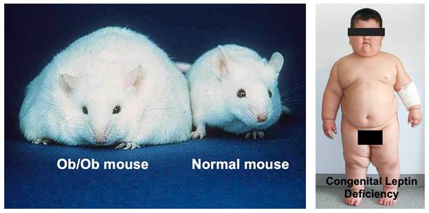
    These children presented with a dramatic phenotype: they were born at normal weight but rapidly developed severe obesity in early childhood, often reaching extreme weights by age 5 or 6. They had insatiable hunger, constantly seeking food, and displayed food-seeking behaviors that disrupted family life. They also had delayed puberty due to hypogonadotropic hypogonadism, leptin’s absence prevented normal sexual maturation.

    The medical implications extended beyond obesity. Many patients with congenital leptin deficiency developed severe metabolic complications:

    Lipodystrophy-like Features: While the genetic form involves too little leptin from too much fat, patients with lipodystrophy, a group of disorders characterized by loss of body fat, also have leptin deficiency, but from the opposite cause: too little fat tissue. Whether from genetic mutations or acquired fat loss, the lack of leptin causes similar metabolic derangements.

    Severe Hypertriglyceridemia: Leptin deficiency causes marked elevations in triglycerides, sometimes reaching levels over 1000 mg/dL or higher. This creates visible lipemia—the blood plasma appears milky white due to excess fat particles. Such extreme elevations dramatically increase the risk of pancreatitis, a potentially life-threatening inflammation of the pancreas.

    Fatty Liver Disease: Without adequate leptin signaling, the liver accumulates excessive fat, leading to hepatic steatosis and potentially progressing to more severe liver damage.

    Insulin Resistance and Diabetes: The metabolic dysfunction extends to glucose metabolism, with many leptin-deficient patients developing insulin resistance and type 2 diabetes at young ages.

    Immune Dysfunction: Leptin plays a role in immune function, and deficient patients often have increased susceptibility to infections.

    Leptin Replacement: A Remarkable Treatment

    When leptin-deficient patients were identified, the obvious treatment approach was leptin replacement therapy—essentially, providing the hormone these patients couldn’t make. The results were nothing short of miraculous.

    Children and adults with congenital leptin deficiency who received recombinant leptin (metreleptin) experienced dramatic transformations. Within weeks to months of starting treatment, patients showed:

    Marked Weight Loss: Patients lost substantial weight, sometimes 50% or more of their body weight over the first year of treatment. The weight loss continued until they reached near-normal weight ranges.

    Normalized Eating Behavior: The constant hunger disappeared. Patients who had been consumed by food-seeking behavior developed normal appetite regulation. Families reported life-changing improvements in quality of life.

    Resolution of Metabolic Abnormalities: Triglyceride levels plummeted from dangerous elevations to normal ranges within weeks. The lipemia resolved. Fatty liver improved. Glucose metabolism normalized.

    Sexual Development: Children with delayed puberty began normal pubertal progression. Adults with hypogonadism experienced normalization of sex hormones.

    Improved Quality of Life: Beyond the physical changes, patients reported profound improvements in well-being, energy, and ability to participate in normal activities.

    The treatment was equally effective in patients with acquired lipodystrophy. Whether from genetic causes, autoimmune destruction of fat tissue, or HIV treatment-related fat loss, patients with severe lipodystrophy and leptin deficiency showed similar dramatic responses to leptin replacement. The FDA approved metreleptin in 2014 specifically for treating complications of leptin deficiency in patients with congenital or acquired lipodystrophy.

    The Broader Impact and Unexpected Discoveries

    The leptin story illustrates both the power and the limitations of translational research. While leptin replacement is spectacularly effective for the rare patients with leptin deficiency, it largely failed as a treatment for common obesity. Most obese people have high leptin levels—their problem isn’t leptin deficiency but leptin resistance, where the brain fails to respond appropriately to the hormone’s signal. This parallels type 2 diabetes, where the problem is usually insulin resistance rather than insulin deficiency.

    Nevertheless, leptin research opened entire new fields of investigation. Scientists discovered that leptin is part of a complex network of hormones and neural circuits regulating energy balance. The hypothalamic pathways that respond to leptin have been mapped in extraordinary detail. We now understand that leptin regulates not just appetite and metabolism but also immunity, reproduction, bone metabolism, and many other physiological systems.

    The discovery validated the concept that body weight is biologically regulated—that the body has sophisticated mechanisms for sensing and defending its energy stores. This shifted the conversation about obesity from one of simple willpower to recognition of underlying biological drives that make weight loss and maintenance challenging.

    Legacy of Discovery

    Jeffrey Friedman’s identification of leptin stands as one of the landmark achievements in metabolism research. The work earned him numerous awards, including the Lasker Award (often called “America’s Nobel”) and the Shaw Prize. More importantly, it provided:

    Understanding of Weight Regulation: Leptin revealed that body weight isn’t simply a matter of calories in versus calories out, but involves active biological regulation through hormonal signaling between fat tissue and the brain.

    Treatment for Rare Diseases: For patients with congenital leptin deficiency or lipodystrophy, leptin replacement is genuinely life-saving, preventing deadly complications like pancreatitis and providing normal quality of life.

    Research Framework: The discovery established paradigms for studying energy homeostasis and identified key neural circuits in the hypothalamus that have become targets for developing new metabolic therapies.

    Validation of Genetic Approaches: The success of positional cloning to identify leptin validated this approach and paved the way for identifying many other disease genes.

    The story of leptin demonstrates how basic research—studying mutant mice in a laboratory—can lead to profound clinical applications. Those ob/ob mice that puzzled researchers in 1949 eventually revealed fundamental truths about how our bodies regulate weight and led to treatments that transformed lives for patients with devastating metabolic disorders.

    The journey from a curious mutation in mice to a life-saving treatment for humans took nearly half a century. It required insights from genetics, molecular biology, endocrinology, and clinical medicine. It stands as a testament to the value of basic research and the unexpected ways that understanding fundamental biology can ultimately benefit human health. The ob mouse, once just an oddity in a research colony, became the key to unlocking one of metabolism’s most important secrets.

  • Does Viagra or Cialis Help Improve Sexual Sensitivity in Women?

    When most people think of Viagra (sildenafil) or Cialis (tadalafil), they associate these medications exclusively with male sexual dysfunction. However, researchers have long been curious about whether these drugs might offer similar benefits for women experiencing sexual difficulties. The answer, as it turns out, is considerably more complex than a simple yes or no.

    Understanding the Pharmacology: How PDE5 Inhibitors Work

    To understand whether these medications could help women, we first need to explore how they work at the biochemical level.

    The Nitric Oxide Pathway

    Both Viagra and Cialis belong to a class of drugs called phosphodiesterase type 5 (PDE5) inhibitors. Their mechanism of action centers on the nitric oxide (NO) signaling pathway, which plays a crucial role in smooth muscle relaxation throughout the body.

    Here’s how the cascade works:

    Step 1: Sexual Stimulation and Nitric Oxide Release When sexual arousal occurs (through physical or psychological stimulation), nerve endings and endothelial cells lining blood vessels release nitric oxide. This is a critical first step that these medications cannot replace or initiate on their own.

    Step 2: Activation of Guanylate Cyclase Nitric oxide binds to and activates an enzyme called guanylate cyclase within smooth muscle cells. This enzyme then converts guanosine triphosphate (GTP) into cyclic guanosine monophosphate (cGMP).

    Step 3: cGMP and Smooth Muscle Relaxation The accumulation of cGMP triggers a series of events that ultimately lower calcium levels within smooth muscle cells. This reduction in intracellular calcium causes the smooth muscles surrounding blood vessels to relax, leading to vasodilation (widening of blood vessels) and increased blood flow to the tissue.

    Step 4: The Role of PDE5 Under normal circumstances, an enzyme called phosphodiesterase type 5 (PDE5) breaks down cGMP, which eventually returns the smooth muscle to its non-relaxed state and reduces blood flow back to baseline levels.

    Step 5: How PDE5 Inhibitors Work Viagra and Cialis work by selectively inhibiting PDE5, preventing it from breaking down cGMP. This allows cGMP to accumulate to higher levels and persist for longer periods, prolonging smooth muscle relaxation and maintaining increased blood flow to the target tissues.

    Key Biochemical Principle

    It’s essential to understand that PDE5 inhibitors do not create arousal or initiate the nitric oxide cascade. They simply amplify and prolong the body’s natural response to sexual stimulation by preventing the breakdown of cGMP. Without the initial release of nitric oxide from arousal, these medications have little to no effect.

    Effects on Muscles and Blood Flow

    The smooth muscle relaxation induced by PDE5 inhibitors affects blood vessels throughout the body, though the medications show some tissue selectivity.

    In Men

    In men, PDE5 is highly concentrated in the corpus cavernosum of the penis. When PDE5 is inhibited, blood flow to this erectile tissue increases dramatically, facilitating and maintaining an erection sufficient for sexual activity. The mechanism is straightforward: increased blood flow leads to engorgement, which produces the physical changes necessary for penetrative intercourse.

    In Women: The Anatomical Parallel

    Women have analogous erectile tissues that respond to increased blood flow during arousal:

    • Clitoral tissue: The clitoris contains erectile tissue that engorges with blood during arousal, similar to penile tissue
    • Vaginal walls: Increased blood flow leads to vaginal lubrication through transudate (fluid passing through vessel walls)
    • Labia: The labia minora and majora also experience increased blood flow and engorgement during arousal

    Theoretically, PDE5 inhibitors should enhance blood flow to these tissues in women just as they do in men, since the underlying biochemical mechanisms are identical. Studies have confirmed that these medications do indeed increase genital blood flow in women.

    The Critical Question: Does Increased Blood Flow Equal Enhanced Sexual Function?

    This is where the science becomes fascinating and reveals a fundamental difference between male and female sexual response.

    What the Research Shows

    Multiple clinical trials have investigated whether Viagra or Cialis improve sexual function in women, particularly those with sexual arousal disorder or hypoactive sexual desire disorder. The results have been largely disappointing:

    Physiological Response vs. Subjective Experience Studies consistently demonstrate that PDE5 inhibitors successfully increase genital blood flow in women. Using devices that measure vaginal pulse amplitude and lubrication, researchers have documented significant increases in these physiological markers of arousal when women take these medications.

    However, when women are asked about their subjective experience—whether they feel more aroused, experience greater pleasure, or find sex more satisfying—the medications typically show no significant benefit compared to placebo.

    The Disconnect: Why Blood Flow Isn’t Enough

    This disconnect between physiological and psychological arousal reveals something important about female sexuality. Unlike the relatively linear relationship between genital blood flow and function in men (more blood flow = firmer erection = ability to have intercourse), female sexual response is considerably more complex.

    Several factors contribute to this complexity:

    1. The Role of Psychological Arousal Female sexual satisfaction appears to depend much more heavily on psychological factors including emotional connection, mental arousal, context, stress levels, relationship quality, and mood. Simply increasing blood flow to genital tissues does not address these psychological components.

    2. Concordance Between Mind and Body Research on sexual concordance (the agreement between physical and mental arousal) shows that women typically have much lower concordance than men. A woman’s body may show physiological signs of arousal while her mind feels unaroused, or vice versa. Men generally show high concordance between genital response and subjective arousal.

    3. Multiple Components of Sexual Response Female sexual dysfunction often involves multiple interrelated factors including desire, arousal, orgasm, pain, and satisfaction. Addressing blood flow alone targets only one small piece of this multifaceted puzzle.

    Specific Conditions Where PDE5 Inhibitors Might Help Women

    While general use in women has not proven beneficial, there are specific situations where these medications may offer some advantage:

    1. Sexual Dysfunction Related to Antidepressants

    Some studies suggest that PDE5 inhibitors may help counteract sexual side effects caused by selective serotonin reuptake inhibitors (SSRIs), though results are mixed. The medications may help improve physiological aspects of arousal that SSRIs impair.

    2. Clitoral Blood Flow Issues

    In cases where reduced clitoral sensation is specifically related to insufficient blood flow (rather than nerve damage or psychological factors), PDE5 inhibitors might theoretically provide benefit. However, this represents a small subset of female sexual dysfunction.

    3. Postmenopausal Women

    Some research has explored whether these medications might help address arousal difficulties in postmenopausal women experiencing changes in genital blood flow, though results remain inconclusive.

    4. Women with Specific Medical Conditions

    Conditions that affect blood flow systemically, such as diabetes or cardiovascular disease, might theoretically benefit from improved genital blood flow, though clinical evidence is limited.

    Safety Considerations for Women

    While Viagra and Cialis are generally well-tolerated, they are not without side effects:

    • Headaches
    • Flushing
    • Nasal congestion
    • Visual disturbances
    • Dizziness
    • Indigestion

    More importantly, these medications are contraindicated in people taking nitrate medications (commonly prescribed for heart conditions) because the combination can cause dangerous drops in blood pressure.

    Additionally, neither Viagra nor Cialis is FDA-approved for use in women. This means that any prescription would be off-label, and insurance typically would not cover the cost.

    What Actually Helps Women with Sexual Dysfunction?

    Given that PDE5 inhibitors generally don’t improve sexual satisfaction in women, what does work?

    1. Addressing Psychological Factors

    Sex therapy, cognitive behavioral therapy, and mindfulness-based approaches have shown significant benefit for many women with sexual difficulties. These therapies address the psychological components that play such a crucial role in female sexuality.

    2. Hormone Therapy

    For women experiencing sexual dysfunction related to hormonal changes (particularly during perimenopause and menopause), hormone replacement therapy or topical estrogen may help address vaginal dryness, pain, and atrophy.

    3. FDA-Approved Medications for Women

    • Flibanserin (Addyi): Approved for premenopausal women with hypoactive sexual desire disorder, this medication works on neurotransmitter systems rather than blood flow
    • Bremelanotide (Vyleesi): An injectable medication also approved for low sexual desire in premenopausal women, working through melanocortin receptors

    4. Treating Underlying Conditions

    Addressing conditions that contribute to sexual dysfunction—such as depression, anxiety, relationship issues, chronic pain, or medication side effects—often proves more effective than attempting to treat the sexual symptoms directly.

    5. Pelvic Floor Physical Therapy

    For women experiencing pain during intercourse or difficulty with arousal related to pelvic floor dysfunction, specialized physical therapy can be remarkably helpful.

    The Bottom Line

    From a purely pharmacological perspective, PDE5 inhibitors like Viagra and Cialis do exactly what they’re designed to do in women: they inhibit the PDE5 enzyme, increase cGMP levels, promote smooth muscle relaxation, and enhance blood flow to genital tissues. The biochemical mechanism is sound, and the physiological effects are measurable.

    However, improved blood flow does not translate to improved sexual sensitivity, pleasure, or satisfaction for most women. This reveals an important truth about human sexuality: the same biochemical pathway can have very different practical outcomes depending on the complex interplay of psychological, emotional, and social factors that influence sexual experience.

    Female sexuality is not simply male sexuality in a different anatomical package. The relative importance of psychological versus physiological factors differs, and effective treatments must address this reality. For women experiencing sexual difficulties, a comprehensive approach that considers the full spectrum of physical, psychological, and relational factors is far more likely to help than a medication designed to address a primarily mechanical problem.

    If you’re experiencing sexual difficulties, the most important first step is having an open conversation with a healthcare provider who can help identify the specific factors contributing to your concerns and develop a tailored treatment approach. While Viagra and Cialis probably won’t be part of that solution for most women, numerous other effective options exist.


    Medical Disclaimer: This article is for informational purposes only and does not constitute medical advice. Always consult with a qualified healthcare provider regarding medical questions, conditions, or treatment options.

  • A Dangerous Cut: How Eliminating Research Funding Threatens Public Health

    Recent federal budget bills and policy decisions propose drastic cuts that would eliminate funding for critical scientific research at the National Institutes of Health (NIH) and various universities across the nation. While fiscal responsibility is important, these specific budgetary and policy decisions are not just penny-wise and pound-foolish; they are a direct threat to the health and well-being of every American.

    NIH

    The NIH is the largest biomedical research agency in the world, responsible for groundbreaking discoveries that have led to treatments for diseases ranging from cancer to Alzheimer’s, and the development of life-saving vaccines. Universities, often in partnership with NIH, are hotbeds of innovation, fostering the next generation of scientists and pushing the boundaries of medical knowledge. Gutting their funding is akin to dismantling our medical lifeline.

    NIH

    The Dire Consequences for Public Health:

    • Stifled Innovation and Delayed Cures: Imagine a future where the next breakthrough in cancer treatment, the vaccine for an emerging pandemic, or a more effective therapy for chronic diseases never sees the light of day. Without sustained funding, researchers cannot conduct the essential studies, trials, and investigations necessary to make these life-changing discoveries. We will simply stop making progress.
    • Brain Drain and Loss of Expertise: Our nation’s top scientists are drawn to environments where their work is supported and valued. If funding evaporates, we risk a significant “brain drain” as talented researchers seek opportunities in other countries that prioritize scientific advancement. This loss of expertise will have long-lasting, detrimental effects on our capacity to address future health challenges.
    • Increased Healthcare Costs: While it may seem counterintuitive, investing in preventative research and new treatments often reduces long-term healthcare costs. By understanding diseases better and developing more effective interventions, we can prevent serious illnesses, reduce hospital stays, and improve quality of life, ultimately saving billions in treatment expenses. Cutting research funding today will likely lead to far greater healthcare burdens tomorrow.
    • Weakened Pandemic Preparedness: The COVID-19 pandemic starkly illustrated the critical importance of robust scientific research for rapid response and vaccine development. Eliminating funding now would leave us dangerously vulnerable to future public health crises, effectively disarming us in the face of unknown pathogens.
    • Erosion of Public Trust and Global Leadership: America has long been a global leader in scientific discovery. Retreating from this commitment sends a troubling message to the world and undermines public trust in the ability of our institutions to protect citizens’ health.

    Who Should Oppose These Moves?

    The potential ramifications of these cuts are too significant to ignore. Everyone with an interest in a healthier future should actively oppose these budgetary decisions. This includes:

    • Patients and Patient Advocates: Those directly affected by diseases and their families, who rely on research for hope and new treatments.
    • Healthcare Professionals: Doctors, nurses, and other medical providers who understand the critical role research plays in advancing clinical care.
    • Scientists and Academics: The researchers themselves, who are on the front lines of discovery, and the institutions that house them.
    • Public Health Organizations: Groups dedicated to improving community health and preventing disease.
    • Concerned Citizens: Anyone who believes in the power of science to improve lives and secure a healthier future for all.

    We cannot afford to mortgage our future health for short-sighted budget cuts. Contact your elected officials, support organizations advocating for scientific research, and make your voice heard. Our collective health depends on it.

  • What Is Epithalon? The Russian Longevity Peptide That Extends Lifespan

    If you’ve gone deep into the longevity and anti-aging rabbit hole, you’ve probably heard whispers about Epithalon (also spelled Epitalon)—a peptide that supposedly extends lifespan by acting on your cells’ internal aging clock. Unlike peptides that improve appearance or body composition, Epithalon claims to address aging at its most fundamental level: the shortening of your telomeres.

    This sounds like science fiction, but Epithalon has over 30 years of Russian research behind it. So what is it really? How does it work? And can a simple four-amino-acid peptide actually slow down aging?

    Let’s break down Epithalon—the peptide that targets the very mechanism of cellular aging.

    The Discovery: Russian Longevity Research

    The story of Epithalon begins in the Soviet Union during the Cold War era, when the USSR was heavily investing in gerontology (the study of aging) and life extension research.

    Professor Vladimir Khavinson and the St. Petersburg Institute

    In the 1970s-1980s, Professor Vladimir Khavinson at the St. Petersburg Institute of Bioregulation and Gerontology became obsessed with one question: What controls the aging process at the most fundamental level?

    His hypothesis was controversial: he believed that the pineal gland—a small, pine-cone-shaped gland in your brain—was the master regulator of aging.

    The Pineal Gland’s Role:

    The pineal gland produces melatonin, which regulates your circadian rhythm (sleep-wake cycle). But Khavinson noticed something interesting: as people age, their pineal gland calcifies and produces less melatonin. This decline correlates strongly with other markers of aging.

    He theorized that the pineal gland produces other regulatory peptides that control aging throughout the body, and that declining pineal function drives the aging process.

    The Extraction and Synthesis

    Khavinson’s team extracted peptides from the pineal glands of young calves. After years of fractionation and testing, they isolated a specific tetrapeptide (four amino acids) that had remarkable effects on lifespan in animal models.

    The sequence:

    Ala-Glu-Asp-Gly (Alanine-Glutamate-Aspartate-Glycine)

    They called it Epithalon (also known as Epithalamin when referring to the natural pineal extract).

    The Results:

    In animal studies spanning three decades, Epithalon demonstrated:

    42% increase in lifespan in aged rats

    27% increase in lifespan in fruit flies

    Extended lifespan in mice by normalizing age-related changes

    Restored reproductive function in old female rats

    Normalized circadian rhythms in aged animals

    These weren’t subtle improvements—these were dramatic extensions of both lifespan and healthspan (years lived in good health).

    The Mechanism: Telomeres and the Hayflick Limit

    To understand how Epithalon works, you need to understand telomeres—the “aging clock” inside every cell.

    What Are Telomeres?

    Imagine your DNA as shoelaces. The plastic tips on the ends of shoelaces (aglets) prevent them from fraying. Telomeres are like those plastic tips—they’re protective caps on the ends of your chromosomes.

    The structure:

    Telomeres are repetitive DNA sequences (TTAGGG repeated thousands of times) that don’t code for any proteins. They’re essentially “junk” DNA whose only job is to protect the important genetic information on your chromosomes.

    The Problem: Telomere Shortening

    Every time a cell divides, the DNA replication machinery can’t quite copy all the way to the very end of the chromosome. A small piece of the telomere is lost with each division.

    The Hayflick Limit:

    In the 1960s, Dr. Leonard Hayflick discovered that normal human cells can only divide about 40-60 times before they stop. This is called the Hayflick Limit.

    Why do they stop? Because their telomeres become critically short.

    What happens when telomeres get too short:

    1. Cellular senescence: The cell stops dividing and becomes a “zombie cell”—it’s alive but no longer functional. These senescent cells secrete inflammatory molecules that damage surrounding tissues.

    2. Apoptosis: The cell dies (programmed cell death)

    3. Genetic instability: If the cell tries to divide with critically short telomeres, chromosomes can fuse together or break, leading to cancer or cell death

    This is why we age: Your cells can only divide a limited number of times. Eventually, you run out of functional cells, and tissues deteriorate.

    The Solution: Telomerase

    There’s an enzyme called telomerase that can add DNA sequences back onto telomeres, essentially “refilling” them.

    Telomerase structure:

    • TERT (Telomerase Reverse Transcriptase): The protein enzyme component
    • TERC (Telomerase RNA Component): The RNA template that guides DNA synthesis

    How it works:

    Telomerase uses its RNA template to add TTAGGG repeats back onto chromosome ends, lengthening telomeres and allowing more cell divisions.

    The catch: Most adult cells DON’T produce telomerase. It’s only active in:

    • Germ cells (sperm and eggs)
    • Stem cells
    • Some immune cells
    • Cancer cells (which is why they’re “immortal”)

    This is your body’s anti-cancer defense mechanism. If all cells had active telomerase, they could divide forever—which is essentially what cancer is.

    But it also means your normal cells have a built-in expiration date.

    How Epithalon Works: Activating Telomerase

    Here’s where Epithalon comes in. Research suggests it can activate telomerase in somatic cells (normal body cells) temporarily and safely.

    The Molecular Mechanism

    Step 1: Pineal Gland Stimulation

    Epithalon appears to act directly on the pineal gland, stimulating it to produce more of its natural regulatory peptides and hormones, particularly melatonin.

    The exact receptor mechanism isn’t fully characterized, but research suggests it binds to specific receptors on pinealocytes (pineal gland cells).

    Step 2: Gene Expression Changes

    Epithalon modulates the expression of genes involved in cellular aging and stress response. Specifically, it appears to:

    • Upregulate TERT gene expression (the telomerase enzyme)
    • Increase production of the telomerase enzyme
    • Enhance the enzyme’s activity

    Step 3: Telomerase Activation

    With more telomerase present and active, cells can add DNA sequences back onto their telomeres. This doesn’t make cells immortal—it just “resets” some of the telomere shortening that has occurred.

    The evidence:

    Studies on human cells in culture showed:

    • Telomere length increased by an average of ~10-15% after Epithalon treatment
    • Cells that had stopped dividing (senescent) could divide again
    • Lifespan of cells in culture extended by 20-40%

    Step 4: Melatonin Normalization

    Epithalon also normalizes melatonin production from the pineal gland. This has cascading effects:

    • Improved circadian rhythm
    • Better sleep quality
    • Enhanced antioxidant defenses (melatonin is a powerful antioxidant)
    • Improved neuroendocrine function

    The Circadian Rhythm Connection

    One of Epithalon’s most immediate effects is on circadian rhythm. The pineal gland releases melatonin in response to darkness, signaling your body that it’s time to sleep.

    As you age:

    • Pineal gland calcifies
    • Melatonin production decreases
    • Circadian rhythm becomes less robust
    • Sleep quality deteriorates

    Epithalon appears to “reset” the pineal gland, restoring more youthful melatonin patterns.

    Why this matters for aging:

    Your circadian rhythm controls hundreds of genes throughout your body. When it’s disrupted:

    • Immune function declines
    • Metabolic health worsens
    • Inflammation increases
    • Cognitive function deteriorates

    By normalizing circadian rhythms, Epithalon may indirectly slow many aspects of aging.

    The Antioxidant and Anti-Inflammatory Effects

    Beyond telomeres and circadian rhythms, Epithalon has systemic effects on oxidative stress and inflammation.

    Reducing Lipid Peroxidation

    Lipid peroxidation is the “rusting” of fats in your body. Free radicals attack lipids in cell membranes, creating chain reactions that damage cells.

    Studies show Epithalon:

    • Reduces lipid peroxidation by 30-40%
    • Increases antioxidant enzyme activity (SOD, catalase, glutathione peroxidase)
    • Protects mitochondria from oxidative damage

    Why this matters:

    Oxidative damage accumulates with age and contributes to cardiovascular disease, neurodegeneration, and general tissue deterioration.

    Normalizing Cortisol

    Epithalon appears to normalize the hypothalamic-pituitary-adrenal (HPA) axis, which controls your stress response.

    In aged animals, Epithalon:

    • Reduced elevated baseline cortisol
    • Improved cortisol response to stress (better stress adaptation)
    • Restored more youthful hormone patterns

    Chronically elevated cortisol accelerates aging, so normalizing this is significant.

    Practical Effects: What Users Report

    Unlike peptides with immediate visible effects (like BPC-157 or GHK-Cu), Epithalon’s benefits are subtle and long-term. Here’s what users typically experience:

    Effect 1: Improved Sleep Quality (Most Common, First to Notice)

    Timeline: Within 3-7 days

    Most users report:

    • Falling asleep more easily
    • Deeper, more restorative sleep
    • More regular sleep-wake cycle
    • Waking up more refreshed

    Why: Melatonin normalization from pineal gland stimulation

    This is often the only immediate, perceptible effect. Everything else is more subtle.

    Effect 2: Enhanced Energy and Vitality

    Timeline: Weeks 2-4

    Users often describe:

    • More consistent energy throughout the day
    • Less afternoon crashes
    • Better stress resilience
    • General sense of “feeling younger”

    Why: Normalized circadian rhythm → better hormone patterns → improved cellular metabolism

    Effect 3: Cognitive Improvements

    Timeline: Weeks 3-6

    Some users report:

    • Improved mental clarity
    • Better memory (especially short-term)
    • Faster cognitive processing
    • Reduced brain fog

    Why: Melatonin’s neuroprotective effects + reduced oxidative stress in brain

    Effect 4: Visual Changes (Subtle)

    Timeline: Weeks 6-10

    Some users notice:

    • Slight improvement in skin quality
    • Reduction in age spots
    • Better hair quality
    • Improved wound healing

    Why: Cellular rejuvenation from telomerase activation + antioxidant effects

    These changes are much more subtle than with peptides like GHK-Cu. Don’t expect dramatic cosmetic changes.

    Effect 5: Biomarker Improvements

    Timeline: Measurable at weeks 8-12

    Studies and anecdotal reports show:

    • Improved lipid profiles (cholesterol, triglycerides)
    • Better glucose metabolism
    • Normalized blood pressure
    • Improved immune markers

    These require blood tests to measure—you won’t “feel” them directly.

    The Dosing Protocol: How Epithalon Is Used

    Epithalon is typically used in short cycles rather than continuously.

    Standard Longevity Protocol

    Dose: 10 mg per day (sometimes split as 5 mg twice daily)

    Duration: 10-20 days

    Frequency: 2-4 times per year

    Route: Subcutaneous injection (same as other peptides)

    Example cycle:

    • 10 mg daily for 10 days
    • Off for 4-6 months
    • Repeat cycle

    Why cycles instead of continuous:

    The theory is that telomerase activation shouldn’t be constant (cancer risk), so pulsing it in short cycles achieves benefits while minimizing risk.

    Intensive Anti-Aging Protocol

    Dose: 10 mg per day

    Duration: 20 days

    Frequency: 4 times per year (once per quarter)

    Example annual schedule:

    • January: 20-day cycle
    • April: 20-day cycle
    • July: 20-day cycle
    • October: 20-day cycle

    This is the protocol most commonly used in Russian longevity clinics.

    Timing of Injections

    Before bed: Most common recommendation

    • Synergizes with natural melatonin production
    • Doesn’t interfere with daytime activities
    • Enhances sleep quality effects

    Twice daily: Some protocols use 5 mg morning + 5 mg evening

    • May provide more consistent effects
    • More hassle (two injections per day)

    Administration

    Route: Subcutaneous injection

    Sites: Abdomen, thighs (same as other peptides)

    Supplies: Insulin syringes (29-31 gauge)

    Reconstitution: 2-2.5 mL bacteriostatic water per 10 mg vial

    Biomarker Tracking: Measuring Epithalon’s Effects

    Because Epithalon’s effects are subtle and long-term, tracking biomarkers is important.

    Telomere Length Testing

    The gold standard for measuring Epithalon’s effect:

    Tests available:

    • TeloYears (mail-in test)
    • SpectraCell Laboratories
    • Life Length (most detailed, expensive)

    Cost: $100-500 depending on test

    Protocol:

    • Baseline test before first Epithalon cycle
    • Re-test 6-12 months later (after 2-4 cycles)
    • Telomere lengthening or slower shortening indicates effectiveness

    Important: Single tests can vary, so trends over time are more meaningful than one-off measurements.

    Other Relevant Biomarkers

    Complete blood panel:

    • Lipids (Total cholesterol, LDL, HDL, Triglycerides)
    • Glucose and HbA1c
    • Inflammatory markers (hsCRP)
    • Liver and kidney function

    Hormones:

    • Cortisol (morning and evening)
    • Melatonin (salivary test)
    • IGF-1 (marker of growth hormone)

    Advanced markers:

    • 8-OHdG (oxidative DNA damage)
    • Advanced Glycation End-products (AGEs)
    • Senescent cell markers (research/clinical only)

    The Cancer Question: Is Telomerase Activation Safe?

    This is the elephant in the room. If cancer cells use telomerase to become immortal, isn’t activating telomerase dangerous?

    The Theoretical Concern

    Cancer cells reactivate telomerase, allowing unlimited cell divisions. This is why they’re “immortal” and can form tumors.

    If you artificially activate telomerase in normal cells, could you increase cancer risk?

    Why Researchers Think Epithalon Is Safe

    Reason 1: Temporary Activation

    Epithalon activates telomerase briefly (10-20 days), not continuously. Cancer requires sustained telomerase activation.

    Reason 2: Normal Cells Have Other Safeguards

    Even with telomerase active, normal cells have multiple anti-cancer mechanisms:

    • p53 (tumor suppressor gene)
    • Cell cycle checkpoints
    • Immune surveillance

    Cancer requires multiple mutations, not just telomerase.

    Reason 3: 30+ Years of Animal Data

    In animal studies, Epithalon didn’t increase cancer rates. In some studies, it actually reduced cancer incidence (possibly by improving immune function).

    Reason 4: Clinical Use in Russia

    Epithalon has been used in Russian clinics for decades without reported increases in cancer.

    The Caveat

    If you already have cancer (even undiagnosed), activating telomerase could theoretically accelerate tumor growth.

    Recommendations:

    • Get appropriate cancer screenings before using Epithalon
    • Don’t use if you have active cancer
    • Monitor for any unusual symptoms
    • Consider it an “insurance policy” for healthy aging, not a treatment

    Epithalon vs. TA-65: The Telomerase Wars

    Epithalon isn’t the only telomerase activator on the market. TA-65 (a compound derived from astragalus root) is another option.

    TA-65 (Astragalus Extract)

    Mechanism: Activates telomerase through different pathway

    Form: Oral capsules (no injections needed)

    Dosing: Daily supplementation (250-1000 mg/day)

    Evidence: Mixed results, some studies show telomere lengthening

    Cost: $200-600/month (expensive)

    Epithalon vs. TA-65 Comparison

    Epithalon advantages:

    • More robust research (30+ years, multiple animal models)
    • More dramatic telomere lengthening in studies
    • Additional benefits (circadian rhythm, melatonin)
    • Cheaper ($50-150 per cycle, 2-4 cycles per year = $100-600/year)

    TA-65 advantages:

    • Oral (no injections)
    • Commercially available and marketed legally
    • Some human clinical trial data
    • Continuous use (not cycles)

    Verdict: Epithalon has more compelling research but requires injections. TA-65 is more convenient but weaker evidence and very expensive.

    The Russian Connection: Pharmaceutical Availability

    Epithalon is registered as a pharmaceutical drug in Russia under the brand name Endoluten.

    In Russia:

    • Prescribed by doctors for age-related conditions
    • Used in longevity clinics
    • Covered by insurance in some cases
    • Extensive clinical use (thousands of patients)

    In the US and Europe:

    • Not FDA-approved
    • Available as “research chemical”
    • Gray area legally
    • No pharmaceutical-grade version available

    This creates quality control issues for Western users.

    Side Effects and Safety Profile

    Epithalon has an excellent safety profile in the research literature.

    Reported Side Effects (Rare)

    Injection-related:

    • Mild injection site discomfort
    • Rare: redness or itching

    Systemic:

    • Temporary drowsiness (from melatonin increase)
    • Vivid dreams (melatonin effect)
    • Headache (rare, first few days)

    No Significant Adverse Events:

    • No reported organ toxicity
    • No hormonal disruption
    • No significant drug interactions
    • No withdrawal or dependency

    Contraindications

    Avoid if you have:

    • Active cancer
    • History of retinoblastoma (rare eye cancer)
    • Autoimmune conditions (theoretical concern)
    • Pregnancy or breastfeeding (insufficient data)

    Who Should Consider Epithalon?

    Ideal Candidates

    You might benefit if you:

    • Are 40+ years old (telomere shortening becomes significant)
    • Are focused on longevity and healthspan, not just appearance
    • Have family history of age-related diseases
    • Want to optimize cellular aging
    • Are willing to commit to long-term protocol (years)
    • Can afford periodic testing (telomere tests)

    Especially valuable for:

    • Serious biohackers focused on lifespan extension
    • People with premature aging concerns
    • Those with poor sleep/circadian rhythm issues
    • Athletes interested in long-term health (not just performance)

    Who Should Wait or Avoid

    Don’t use if you:

    • Are under 30 (telomeres still long, pineal gland functional)
    • Have active cancer or history of cancer
    • Want immediate, visible results (wrong peptide for that)
    • Are unwilling to track biomarkers (how else will you know it’s working?)
    • Can’t commit to long-term approach

    The Bottom Line: Is Epithalon the Real Deal?

    Epithalon is one of the most scientifically intriguing anti-aging compounds available, but it’s also the hardest to evaluate subjectively.

    What the science shows:

    • Activates telomerase and lengthens telomeres (in vitro and animal studies)
    • Extends lifespan in multiple animal models (20-40%)
    • Normalizes circadian rhythm and melatonin production
    • Reduces oxidative stress and inflammation
    • 30+ years of Russian research and clinical use

    What we don’t have:

    • Large-scale human randomized controlled trials
    • Long-term safety data in Western populations
    • FDA approval or pharmaceutical-grade sources
    • Direct evidence of human lifespan extension

    The value proposition:

    If you’re serious about longevity—actual extension of lifespan, not just looking good—Epithalon is one of the most compelling interventions available.

    At $100-600 per year, it’s far cheaper than most longevity interventions. The risk appears low based on decades of use, and the potential upside (years added to your life) is enormous.

    But this is a long game. You won’t see dramatic effects in weeks or months. This is about influencing the rate at which you age over years and decades.

    For those playing the longevity game seriously, Epithalon deserves strong consideration. For those looking for quick results or cosmetic benefits, there are better options (like GHK-Cu or the Glow Stack).

    Epithalon targets the aging clock itself. Whether it truly extends human lifespan remains to be proven definitively, but the evidence suggests it’s worth the bet.

  • Beyond Tanning: The Biological Operating System Hack of Melanotan 2

    Introduction: More Than Meets the Eye

    When most people hear “Melanotan 2,” they think of a tanning peptide. Some are aware it can enhance libido. But this understanding is like saying water is only for drinking—you’re missing everything else it does. Melanotan 2 (MT2) is actually a synthetic analog of alpha-melanocyte stimulating hormone (α-MSH), a regulatory neuropeptide signaling molecule that interfaces with one of the body’s most fundamental biological control systems: the melanocortin system.

    What makes MT2 fascinating isn’t what it does to your skin. It’s what it does to the underlying biological architecture that separates health from disease. For biohackers and those interested in longevity and wellness, understanding MT2 requires understanding how the melanocortin system functions as a master regulatory network controlling inflammation, metabolism, and energy production throughout your entire body.

    Understanding Alpha-MSH and the Melanocortin System

    The Genesis of Alpha-MSH

    Understanding Alpha-MSH and the Melanocortin System

    Cleavage of POMC by PCs (Prohormone Convertases)

    Alpha-MSH originates from pro-opiomelanocortin (POMC) neurons, primarily located in the arcuate nucleus of the hypothalamus. POMC is cleaved by prohormone convertases to produce several bioactive peptides, including:

    • Alpha-MSH (α-MSH)
    • Beta-MSH (β-MSH)
    • Adrenocorticotropic hormone (ACTH)
    • Beta-endorphin

    Alpha-MSH itself is a tridecapeptide (13 amino acids) derived from ACTH. The native pathway begins when POMC neurons detect signals related to energy balance, immune activation, or inflammatory status. Upon activation, these neurons release α-MSH into various brain regions and the bloodstream, where it acts as a signaling molecule across multiple organ systems.

    The Melanocortin Receptors: A Distributed Network

    Here’s where the biology becomes profound. Alpha-MSH and its analogs like MT2 don’t just bind to melanocytes in your skin. They bind to melanocortin receptors (MC1R through MC5R) distributed throughout your entire body:

    MC1R – Found on melanocytes (skin pigmentation), also expressed on immune cells
    MC3R – Primarily in adipose tissue and hypothalamus (energy homeostasis, inflammation)
    MC4R – Hypothalamus, brainstem, throughout CNS (appetite regulation, energy expenditure, sexual function)
    MC5R – Widely distributed in peripheral tissues including sebaceous glands and immune cells

    The critical insight from research at institutions like the University of Arizona (2011) was discovering that these receptors are located precisely where human biology encounters inflammatory problems or metabolic dysfunction. They’re in endothelial cells, kidneys, immune cells, myocardium, the brain’s reward centers, digestive system, and metabolic tissues.

    This isn’t coincidental; it’s architectural. The melanocortin system evolved as a master regulatory system for coordinating responses across multiple physiological domains simultaneously.

    The Three Biological Failures: A Framework for Understanding Disease

    Modern chronic diseases—type 2 diabetes, cardiovascular disease, neurodegeneration, autoimmune conditions—all converge through three fundamental biological failure points:

    1. Systemic Inflammation – Chronic inappropriate activation of inflammatory cascades
    2. Metabolic Dysfunction – Insulin resistance and impaired cellular energy metabolism
    3. Energy Failure – Mitochondrial dysfunction and insufficient ATP production

    What makes MT2 scientifically compelling is that through melanocortin receptor optimization, it addresses all three simultaneously. Your body isn’t compartmentalized; it’s a unified electromagnetic biochemical information system. When you understand that MT2 activates melanocortin receptors across multiple organ systems at once, you realize it’s not about treating isolated symptoms but optimizing fundamental biological systems.

    Systemic Inflammation: The Master Switch

    The Inflammatory Cascade

    In the modern environment, processed foods, endotoxin exposure, hyperglycemia, oxidative stress, circadian disruption, our immune systems exist in a state of chronic, inappropriately high activation. Macrophages are hypervigilant, dendritic cells are overstimulated, and the T-helper cell TH1/TH17 populations are expanded. This creates a state of smoldering inflammation that slowly damages tissue integrity across all organ systems.

    The key inflammatory mediators include:

    • TNF-α (Tumor Necrosis Factor alpha) – Pro-inflammatory cytokine driving insulin resistance
    • IL-6 (Interleukin-6) – Acute phase protein associated with chronic inflammation
    • IL-1β (Interleukin-1 beta) – Inflammatory cytokine activating NFκB pathway
    • IL-17 – TH17-secreted cytokine linked to autoimmune conditions

    NFκB (Nuclear Factor kappa B) is the master transcription factor that amplifies this inflammatory cascade. When activated, it triggers production of more inflammatory cytokines, creating a self-perpetuating cycle.

    How MT2 Suppresses Inflammation

    MT2’s anti-inflammatory mechanism operates through several pathways:

    POMC Neuron Activation: Research from Stanford University (2015) demonstrated that POMC neuron activation specifically suppresses NFκB in peripheral tissues. By releasing α-MSH directly into the hypothalamic-pituitary-adrenal (HPA) axis, MT2 essentially turns off the biological switch that drives 95% of chronic disease processes.

    Immune Cell Modulation: MT2 stimulates MC3R and MC4R on macrophages and dendritic cells, shifting the immune response from a pro-inflammatory (TH1/TH17-dominant) state to an anti-inflammatory, regulatory (TH2/T-reg-dominant) state.

    Direct Cytokine Suppression: A study in the 2012 Journal of Immunology study showed that melanocortin stimulation directly inhibits TNF-α, IL-6, and IL-1β production in activated macrophages. A 2014 University of Washington study demonstrated that α-MSH-mediated MC4R signaling decreased IL-6, TNF-α, and IL-17 production by approximately 70% in human blood mononuclear cells; not rat studies, but human data.

    This isn’t symptomatic suppression. You’re biochemically rebalancing the immune system toward tolerance and away from chronic violent activation.

    Metabolic Dysfunction: Restoring Insulin Sensitivity

    The Reality of Insulin Resistance

    Insulin resistance isn’t about insufficient insulin production; Rather, it’s about cells becoming deaf to insulin signaling. This happens through multiple mechanisms:

    1. Inflammatory Damage to IRS-1: Chronic inflammation damages insulin receptor substrate-1 (IRS-1) signaling in muscle cells, preventing insulin from activating glucose uptake pathways
    2. Lipid Accumulation: Excess fatty acid accumulation in mitochondria impairs glucose oxidation, creating a metabolic traffic jam
    3. AGE Formation: Chronic hyperglycemia drives formation of advanced glycation end products (AGEs), which activate RAGE receptors and trigger NFκB, perpetuating inflammation
    4. Mitochondrial Dysfunction: Reduced ATP production prevents the energy-dependent steps of glucose metabolism from functioning properly

    MT2’s Multi-Mechanism Metabolic Restoration

    Mechanism 1 – Inflammatory Resolution: By suppressing systemic inflammation, MT2 reduces SOCS (suppressor of cytokine signaling) proteins that phosphorylate and inactivate IRS-1. Your insulin signal becomes cleaner and more effective at the receptor level.

    Mechanism 2 – Enhanced Fat Oxidation: MT2 stimulates MC4R in the sympathetic nervous system, increasing metabolic rate and fatty acid oxidation. This reduces intramuscular lipid accumulation, allowing mitochondria to process glucose more efficiently.

    Mechanism 3 – Reduced AGE Formation: By reducing NFκB activation, MT2 decreases AGE formation and RAGE signaling, improving glucose metabolism directly.

    Mechanism 4 – AMPK Activation: Research from the Journal of Molecular Endocrinology (2013) demonstrated that melanocortin pathway activation increases AMPK (AMP-activated protein kinase) phosphorylation in skeletal muscle. AMPK is the cellular energy sensor that tells mitochondria to produce more ATP and metabolize glucose more efficiently.

    The result isn’t just lower blood sugar—it’s fundamentally improved cellular responsiveness to insulin at the molecular level. Duke University (2015) showed that POMC neuron activation increases metabolic rate by 22% through sympathetic-mediated increases in brown adipose tissue activity and mitochondrial uncoupling protein expression.

    Energy Production: The Mitochondrial Foundation

    The Invisible Pandemic

    Mitochondrial dysfunction is the invisible pandemic underlying most chronic disease. Modern life—high-carbohydrate processed foods, circadian disruption, chronic stress, toxin exposure—systematically damages mitochondrial function. When mitochondria are impaired, everything breaks:

    • Insufficient ATP for synaptic transmission and memory consolidation
    • Impaired myelin maintenance in neurons
    • Reduced protein synthesis capacity
    • Inability to maintain cellular ion gradients
    • Compromised detoxification pathways

    The symptoms manifest differently depending on which tissues are most affected, but the root cause is the same: energy failure.

    MT2’s Mitochondrial Optimization

    Sympathetic Activation: MT2 increases metabolic rate and upregulates mitochondrial biogenesis through PGC-1α (peroxisome proliferator-activated receptor gamma coactivator 1-alpha) activation. You’re literally creating more and healthier mitochondrial power plants.

    Reduced Oxidative Stress: By decreasing inflammation, MT2 reduces production of mitochondrial-damaging reactive oxygen species (ROS) and pro-inflammatory cytokines that downregulate mitochondrial proteins. Research suggests this can reduce the normal ~60% ATP deficit seen in inflammatory states.

    Improved Substrate Utilization: MT2’s enhancement of insulin signaling allows for better glucose oxidation within mitochondria, providing optimal fuel for the electron transport chain and ATP synthesis.

    System-by-System Analysis

    Cardiovascular System: Endothelial Regeneration

    Your endothelium, the single-cell layer lining your arteries,is responsible for:

    • Nitric oxide (NO) production and vasodilation
    • Anti-coagulation and anti-inflammation
    • Vascular tone regulation
    • Filtering inflammatory molecules

    Endothelial dysfunction occurs through chronic inflammation (inflammatory molecule penetration), oxidative stress (reduced NO bioavailability), and insulin resistance (impaired endothelial mitochondrial function). This leads to oxidized LDL particles activating scavenger receptors on macrophages, foam cell formation, and atherosclerosis.

    MT2’s Cardiovascular Benefits:

    • Suppresses systemic inflammation (reduced TNF-α, IL-6, IL-17)
    • Improves NO production in endothelial cells
    • Increases cyclic GMP in smooth muscle cells
    • Reduces baseline sympathetic tone through cortisol and norepinephrine normalization

    A study in Circulation Research (2011) showed that melanocortin agonism improved endothelial function and reduced atherosclerotic plaque formation. Your cardiovascular system regenerates when inflammation decreases and mitochondrial function improves.

    Renal System: Protecting Filtration

    Kidneys are highly metabolically active organs with massive energy requirements. Kidney disease progresses through:

    • Glomerular endothelial dysfunction causing mesangial cell proliferation
    • Chronic inflammation infiltrating kidney tissue
    • Mitochondrial dysfunction impairing tubular reabsorption
    • Oxidative stress damaging podocyte foot processes

    MT2’s Renal Protection:

    • Endothelial repair through improved NO production
    • Reduced glomerular inflammation preventing mesangial proliferation
    • Enhanced mitochondrial function in renal tubules
    • Improved reabsorption of filtered glucose and amino acids

    A study in the American Journal of Nephrology (2014) demonstrated that melanocortin pathway activation reduced proteinuria and improved glomerular filtration rate by 57% in diabetic nephropathy models.

    Nervous System: Neuroprotection and Neuroregeneration

    The brain is approximately 60% lipids, requires constant ATP, has limited blood supply, and is exquisitely sensitive to inflammation. When systemic inflammation is present, microglial cells (the brain’s immune cells) enter a state of chronic hyperactivation.

    Neuroinflammatory Damage:

    • Activated microglia release TNF-α, IL-6, IL-1β onto neural tissue
    • These cytokines damage synaptic connections, impairing long-term potentiation
    • Promotion of amyloid-beta aggregation (foundational to Alzheimer’s)
    • Reduced brain-derived neurotrophic factor (BDNF)
    • Increased oxidative stress

    MT2’s Neuroprotective Mechanisms:

    Anti-inflammatory Effects: MT2 crosses the blood-brain barrier and suppresses microglial activation through melanocortin receptor signaling. The Journal of Neuroinflammation (2012) showed that α-MSH decreased microglial TNF-α and IL-6 production by approximately 70%.

    Enhanced ATP Production: By improving mitochondrial function systemwide, MT2 ensures the brain has sufficient energy for optimal neurotransmission and synaptic plasticity.

    Insulin Sensitivity: By improving systemic insulin sensitivity, MT2 enhances neuronal glucose uptake and metabolism, critical for cognitive function.

    Applications to Neurodegenerative Disease

    Alzheimer’s Disease: Begins with neuroinflammation and amyloid-beta accumulation creating a vicious cycle. MT2 suppresses microglial activation, reduces neuroinflammation, and reduces amyloid-beta-induced inflammatory amplification. Neurobiology of Aging (2013) showed MT2 reduced amyloid-beta-induced microglial TNF-α production.

    Parkinson’s Disease: Characterized by selective dopaminergic neuron loss in the substantia nigra, driven by neuroinflammation and mitochondrial dysfunction. MT2 suppresses the neuroinflammation damaging dopaminergic neurons, improves mitochondrial function in remaining neurons, and reduces alpha-synuclein aggregation downstream.

    Multiple Sclerosis: An autoimmune condition where the immune system attacks myelin, driven by TH17 overactivity and insufficient T-regulatory cell activity. MT2 shifts the immune response from TH1/TH17 toward TH2/T-reg, reducing the myelin-directed attack and promoting remyelination by oligodendrocytes. The Journal of Neuroinflammation (2014) showed melanocortin agonism increased regulatory T cells and decreased TH17 activity in MS.

    Sexual Function: The Complete Biological Picture

    Erectile dysfunction and low libido are fundamentally about impaired biological systems, not pharmaceutical deficiencies.

    The Requirements for Proper Sexual Function:

    1. Cavernosal smooth muscle relaxation (blood fills corpus cavernosum)
    2. Penile arterial vasodilation (blood flows in)
    3. Central arousal signals from the brain (system activation)

    Where Modern Men Break Down:

    • Chronic stress: Elevated cortisol and norepinephrine create sympathetic dominance—the penis is stuck in fight-or-flight mode, unable to relax smooth muscle
    • Chronic inflammation: Elevated TNF-α, IL-6, IL-17 cause endothelial dysfunction, reduced nitric oxide production, and impaired vasodilation
    • Metabolic dysfunction: Reduced ATP in smooth muscle cells means insufficient energy to maintain the relaxation required for erection

    MT2’s Multi-Level Sexual Enhancement:

    Brain Level: MT2 stimulates melanocortin receptors in the medial preoptic area (MPOA) and paraventricular nucleus (PVN)—the brain’s sexual arousal centers. University of Amsterdam research (2007) showed direct melanocortin agonism in the PVN increases penile erection frequency and intensity by 47%. MT2 also increases dopamine and oxytocin in these regions, fundamental to sexual motivation and desire.

    Endothelial Level: MT2 suppresses systemic inflammation, improves NO production in endothelial cells, and increases cyclic GMP in cavernosal smooth muscle cells, the biological cascade for vasodilation and blood flow.

    Sympathetic Level: By suppressing systemic inflammation (the chronic stressor), MT2 reduces baseline norepinephrine and cortisol, normalizing sympathetic nervous system tone and releasing the fight-or-flight grip on genital tissue.

    Mitochondrial Level: By improving ATP production in smooth muscle cells, MT2 ensures sufficient energy to maintain the metabolic state that smooth muscle relaxation requires.

    The critical distinction: The International Journal of Impotence Research (2009) showed melanocortin agonism produces durable improvements in erectile function long after discontinuation. You’re not masking symptoms, you’re fixing the underlying biology.

    Clinical Considerations and Dosing Principles

    Understanding the Holistic Effect

    A crucial principle: When you take MT2, you don’t get to choose which melanocortin receptors it activates. Unlike targeted drugs that force specific responses, peptides work through your body’s existing regulatory pathways. MT2 activates the entire melanocortin system, you receive all the effects simultaneously.

    This is fundamentally different from pharmaceutical drugs. Drugs act like wrecking balls, forcing your body to respond even against its natural regulatory mechanisms. Peptides can only tell your body to do what it’s already designed to do—they make these natural processes work orders of magnitude better but cannot force unnatural responses.

    The Integration Approach

    MT2 is not a magic bullet or a supplement to add to an existing stack without consideration. The most effective approach:

    1. Address Foundation First: Optimize diet, sleep, circadian rhythm, and movement patterns
    2. Fix Obvious Deficiencies: Address vitamin D, magnesium, essential fatty acids
    3. Strategic Peptide Integration: Introduce MT2 as part of a comprehensive approach to biological optimization
    4. Monitor and Adjust: Track inflammatory markers (hsCRP), metabolic markers (fasting insulin, HbA1c), and functional outcomes

    Dosing Framework

    Typical research dosing ranges from 0.25mg to 1mg, but individual response varies significantly based on:

    • Baseline melanocortin receptor sensitivity
    • Current inflammatory status
    • Metabolic health status
    • Body composition

    Start low (0.25mg) and assess response over 2-3 weeks before adjusting. The goal isn’t to maximize tanning or achieve a specific aesthetic outcome—it’s to optimize the melanocortin system for comprehensive health benefits.

    Broader Implications: A Systems Biology Perspective

    Why This Matters for Longevity

    The aging process is fundamentally characterized by:

    • Progressive accumulation of inflammatory damage
    • Declining mitochondrial function and biogenesis
    • Increasing insulin resistance and metabolic inflexibility
    • Loss of regenerative capacity

    MT2 addresses each of these aging hallmarks through melanocortin system optimization. By maintaining proper inflammatory regulation, metabolic function, and energy production, you’re not just treating age-related diseases, you’re intervening in the aging process itself.

    The Paradigm Shift

    Modern medicine compartmentalizes: dermatologists for skin, cardiologists for heart, neurologists for brain. But your body doesn’t recognize these arbitrary divisions. It’s a unified system where dysfunction in one area cascades to affect all others.

    The melanocortin system represents a biological master controller coordinating responses across these arbitrary medical specialties. Understanding and optimizing this system requires thinking in terms of:

    • Systems biology rather than reductionist medicine
    • Root cause resolution rather than symptom management
    • Biological optimization rather than pharmaceutical intervention

    Research Directions

    Current research is exploring melanocortin receptor targeting for:

    • Type 2 diabetes and metabolic syndrome
    • Cardiovascular disease and atherosclerosis
    • Neurodegenerative diseases
    • Autoimmune conditions
    • Chronic kidney disease
    • Cancer-related cachexia

    The National Institutes of Health (2013) confirmed that the melanocortin pathway is fundamentally involved in regulating systemic inflammation through POMC neurons, metabolic homeostasis through HPA axis regulation, immune function through cytokine modulation, sexual function through brain receptor activation, cardiovascular tone through endothelial signaling, and cognitive function through hippocampal and prefrontal cortex expression.

    Conclusion: Beyond the Surface

    Melanotan 2 is not a tanning peptide with some side benefits. It’s not primarily a libido enhancer. It’s an intervention into one of the body’s fundamental regulatory systems, the melanocortin system that controls the three biological levers separating health from disease.

    Every chronic disease—cardiovascular disease, type 2 diabetes, Alzheimer’s, Parkinson’s, multiple sclerosis, kidney disease, erectile dysfunction, cognitive decline—bottlenecks through the same three mechanisms: systemic inflammation, metabolic dysfunction, and energy failure. MT2, through its activation of melanocortin receptors across multiple organ systems, addresses all three simultaneously.

    This is what happens when you stop treating symptoms and start optimizing fundamental biological systems. When MC4 receptors optimize sympathetic tone and metabolic rate, when AMPK activates in muscle tissue restoring insulin sensitivity, when POMC neurons suppress NFκB and TNF-α throughout the body, when macrophages shift from pro-inflammatory to anti-inflammatory phenotypes, when mitochondria regenerate and produce more ATP with less oxidative stress—you’re not treating a disease. You’re fixing the biological architecture that allows disease to exist.

    For biohackers and those serious about longevity, understanding MT2 means understanding that health is not the absence of disease but the presence of optimized biological function. The melanocortin system is the master lever. MT2 is the tool to optimize it.

    The question isn’t whether MT2 can tan your skin or enhance libido. The question is: what happens when you solve the system that regulates inflammation, metabolism, and energy production across your entire body? The answer is written in the research, confirmed by the biochemistry, and increasingly validated by clinical outcomes.

    This is the future of medicine—not managing symptoms with pharmaceuticals, but optimizing biological systems with our own regulatory molecules. For those willing to understand the science, MT2 represents a glimpse into what’s possible when we work with biology rather than against it.


    Note: This article presents scientific research and biological mechanisms for educational purposes. MT2 is a research peptide, and any therapeutic use should be undertaken with qualified medical supervision. Individual responses vary based on health status, genetics, and other factors. The information presented is for educational purposes and should not be construed as medical advice.

  • The Truth About Conversion Therapy: A History of Harm

    For decades, thousands of LGBTQ individuals were subjected to treatments designed to change their sexual orientation or gender identity. Known as conversion therapy, reparative therapy, or ex-gay therapy, these practices represent one of the darker chapters in modern mental health care. Understanding this history is crucial to preventing its continuation and protecting vulnerable LGBTQ youth today.

    What Is Conversion Therapy?

    Conversion therapy encompasses a range of practices aimed at changing, suppressing, or “curing” a person’s sexual orientation or gender identity. These interventions have taken many forms over the years—from psychoanalysis and behavioral conditioning to religious counseling and, in extreme historical cases, aversive techniques including electroshock therapy and nausea-inducing drugs. The underlying premise has always been the same: that being gay, lesbian, bisexual, or transgender is a disorder that can and should be treated.

    Practitioners have employed various methods, including talk therapy sessions focused on identifying supposed childhood trauma or family dysfunction that “caused” homosexuality, behavioral modification techniques, prayer and religious intervention, and group therapy designed to reinforce heterosexual identity. Some approaches were clinical, conducted by licensed therapists, while others operated within religious organizations with little to no mental health training.

    Gay Conversion Therapy.  Harmful.

    The Origins and Rise of Conversion Therapy

    The concept of homosexuality as a treatable condition emerged from early 20th-century psychiatry, when same-sex attraction was classified as a mental illness. Sigmund Freud himself had a complex view on the matter, while he considered homosexuality a developmental arrest, he famously wrote to a mother in 1935 that it “is nothing to be ashamed of, no vice, no degradation” and expressed skepticism about conversion efforts.

    Sigmund Freud.

    Nevertheless, his followers were less cautious. Post-World War II America saw an intensification of efforts to “cure” homosexuality. Psychoanalysts claimed they could redirect sexual orientation through long-term therapy exploring family relationships and childhood experiences. The 1950s and 1960s brought increasingly aggressive approaches, including aversion therapy—shocking patients or inducing vomiting while showing them same-sex imagery.

    Conversion therapy reached its mainstream peak in the 1970s and early 1980s, paradoxically just as the gay rights movement was gaining momentum. Even after the American Psychiatric Association removed homosexuality from its Diagnostic and Statistical Manual of Mental Disorders in 1973, many practitioners continued offering these treatments, now framing them as helping people whose sexuality conflicted with their religious values or personal desires.

    Promised Results Versus Reality

    Proponents of conversion therapy made bold claims. They reported success rates ranging from 30% to 70%, asserting that motivated individuals could achieve heterosexuality through dedicated treatment. Organizations published testimonials from supposedly “ex-gay” individuals who claimed to have successfully changed their orientation and found happiness in heterosexual marriages.

    The reality told a starkly different story. Rigorous scientific examination revealed that these claimed successes were largely illusory. Most studies supporting conversion therapy suffered from severe methodological flaws: they lacked control groups, relied on self-reporting without objective measures, defined “success” vaguely (often meaning merely behavioral change or suppression rather than orientation change), and failed to conduct long-term follow-up.

    When researchers looked more closely, they found that many “success” stories involved people who had simply learned to suppress their attractions or who were bisexual to begin with. The claimed changes typically proved temporary. Numerous individuals who had been touted as conversion therapy success stories later came forward to acknowledge they remained gay and had only learned to hide or deny their true selves.

    The recidivism rate was devastating. Studies that did conduct follow-ups found that the vast majority of people who underwent conversion therapy eventually acknowledged that their sexual orientation had not changed. Many who initially claimed success later returned to living openly as LGBTQ individuals, often after years of internal struggle and failed attempts to maintain a heterosexual identity.

    The Harm Caused

    Far from being merely ineffective, conversion therapy proved actively harmful. Research documented serious negative outcomes including depression, anxiety, decreased self-esteem, substance abuse, homelessness (particularly among youth rejected by families), social isolation, difficulty with intimacy and relationships, post-traumatic stress disorder, and suicidal ideation and attempts.

    The psychological damage stemmed from the therapy’s fundamental premise: that something core to a person’s identity is broken and shameful. Participants internalized the message that they were defective, leading to profound shame and self-hatred. Many described feeling they had failed when the therapy didn’t “work,” adding guilt to their existing distress. The process of trying to suppress or deny one’s authentic self created lasting psychological wounds.

    For minors subjected to conversion therapy—often at their parents’ insistence—the harm was particularly acute. Young people reported feeling betrayed by caregivers, isolated from peers, and fundamentally broken. Some were sent to residential programs that combined conversion therapy with other abusive practices. The suicide rate among LGBTQ youth who underwent conversion therapy was found to be significantly elevated compared to those who did not.

    Exodus International and the Ex-Gay Movement

    No organization embodied the conversion therapy movement more than Exodus International. Founded in 1976, Exodus became the largest ex-gay ministry in the world, with affiliated churches and counseling centers across North America and beyond. The organization promoted the message that through Christian faith and therapy, people could leave homosexuality behind.

    Exodus leaders appeared on television, published books, and lobbied against LGBTQ rights legislation, citing their existence as proof that being gay was a choice. They offered hope to religious families struggling to reconcile their faith with their children’s sexual orientation, and to LGBTQ individuals tormented by religious teachings that condemned them.

    The scandals that eventually surrounded Exodus were numerous and devastating. Multiple Exodus leaders and spokespeople who had claimed to be “ex-gay” were later discovered in same-sex relationships. John Paulk, once the poster child for the ex-gay movement whose story was featured in national advertising campaigns, eventually acknowledged he was still gay and renounced conversion therapy. Michael Bussee, a co-founder of Exodus, left the organization in 1979, came out as gay, and became a vocal critic of the movement.

    Micharl Bussee, Exodus International Co-Founder who is now Gay.

    In 2012, Alan Chambers, then president of Exodus International, made a stunning admission: the majority of people who went through Exodus programs had not experienced a change in their sexual orientation. He acknowledged the harm the organization had caused. In June 2013, Chambers issued a formal apology to the LGBTQ community and announced that Exodus International would shut down. “I am sorry for the pain and hurt many of you have experienced,” he wrote. “I am sorry that some of you spent years working through the shame and guilt you felt when your attractions didn’t change.”

    The closure of Exodus represented a watershed moment, but it did not end conversion therapy. Many former Exodus affiliates continued operating independently, and new organizations emerged to fill the void.

    Professional Opposition and Scientific Consensus

    As evidence of harm mounted, professional mental health organizations took increasingly firm stands against conversion therapy. The American Psychiatric Association led the way, stating in 2000 that conversion therapy can cause depression, anxiety, and self-destructive behavior. Their position paper emphasized that homosexuality is not a mental disorder and does not require treatment.

    The American Psychological Association conducted a comprehensive review of research in 2009, concluding that conversion therapy was unlikely to be successful and carried significant risk of harm. They advised mental health professionals to avoid telling clients they could change their sexual orientation through therapy. The American Medical Association, American Academy of Pediatrics, American Counseling Association, National Association of Social Workers, and numerous other professional organizations issued similar statements.

    These organizations affirmed that sexual orientation is not a choice and is not changeable through therapeutic intervention. They emphasized that the appropriate therapeutic response to a client struggling with their sexual orientation is to help them accept themselves, not to attempt to change them. The scientific consensus became clear and overwhelming: conversion therapy is neither ethical nor effective.

    Legislative Battles

    The fight over conversion therapy moved into legislative arenas as LGBTQ advocates sought legal protections, particularly for minors. California became the first state to ban conversion therapy for minors in 2012, followed quickly by New Jersey in 2013. These laws prohibited licensed mental health professionals from practicing conversion therapy on patients under 18.

    The legislative efforts sparked fierce opposition. Religious organizations and conservative groups argued that such bans violated parental rights, religious freedom, and free speech. Legal challenges were mounted in multiple states, with opponents claiming that conversion therapy bans prevented therapists from helping clients who voluntarily sought to change their orientation.

    Courts generally upheld the bans. In 2014, the Ninth Circuit Court of Appeals ruled that California’s law was constitutional, finding that the state had a legitimate interest in protecting minors from harmful practices. The Supreme Court declined to hear appeals, letting the ruling stand.

    The momentum built gradually but persistently. By 2025, numerous states, the District of Columbia, and Puerto Rico had enacted similar protections. Many municipalities in states without statewide bans passed local ordinances. The bans typically applied only to licensed professionals working with minors, leaving religious counseling and services for adults largely unregulated, a compromise that allowed passage while leaving some activists unsatisfied.

    However, the battle was far from one-sided. Some states introduced “anti-ban” legislation seeking to protect the practice of conversion therapy as a form of religious or therapeutic freedom. These efforts reflected ongoing cultural divisions over LGBTQ rights and the role of government in regulating healthcare and religious practices.

    Where Things Stand Today

    Conversion therapy exists in a state of contested territory. While banned for minors by licensed professionals in many jurisdictions, it continues in various forms: unlicensed religious counseling, life coaching that falls outside professional regulation, practices in states without protective legislation, and services for adults who voluntarily seek them.

    The professional mental health community remains united in opposition. Major therapeutic organizations continue to condemn the practice and call for comprehensive bans. Research continues to document harm, with particular concern for vulnerable populations including LGBTQ youth, people in religious communities that condemn homosexuality, and individuals in countries where being LGBTQ is criminalized.

    Internationally, the picture is mixed. Some countries, including Brazil, Ecuador, and Malta, have banned conversion therapy. The United Nations and World Health Organization have called for global bans. However, in many parts of the world, conversion therapy not only continues but thrives, often with government support or in societies where LGBTQ identities remain deeply stigmatized.

    The legacy of conversion therapy serves as a sobering reminder of how prejudice can masquerade as treatment and how good intentions—whether from families or even some practitioners—can enable profound harm when built on flawed premises. For LGBTQ individuals, particularly those who survived these interventions, the scars often remain.

    Moving forward, the focus has shifted toward affirmative therapy—approaches that help LGBTQ individuals accept themselves, cope with minority stress and discrimination, and build authentic lives. The question is no longer whether sexual orientation can be changed, but how to support people in thriving as their authentic selves.

    The story of conversion therapy is ultimately about the human cost of treating identity as pathology. It stands as a cautionary tale for mental health care and a reminder that scientific evidence and human dignity must guide therapeutic practice, not cultural prejudice or wishful thinking.

  • The Forgotten Pioneers: How Freedom House Created Modern Emergency Care—Then Vanished

    In 1967, Pittsburgh’s Hill District was a community abandoned by the systems meant to protect it. When residents suffered heart attacks, accidents, or medical emergencies, they often waited in vain for help that never arrived with adequate care. The predominantly Black and impoverished neighborhood had been systematically ignored by city services, including emergency medical transport. But from this injustice emerged a revolution that would transform emergency medicine across America—only to be deliberately erased from history.

    Freedom House Ambulance Service began as a bold experiment in community empowerment and medical innovation. Founded by the Maurice Falk Fund president Phil Hallen and Dr. Peter Safar—whose own daughter had died after an asthma attack during inadequate emergency transport—the service recruited twenty-five Black men from the Hill District. Most were unemployed, some were Vietnam veterans struggling with addiction, and many had been labeled “unemployable” by city welfare offices. They were the kind of men society had written off.

    What happened next was extraordinary. Under Dr. Safar’s guidance, these recruits underwent an intensive 32-week, 300-hour training program covering anatomy, physiology, CPR, advanced first aid, nursing, and defensive driving. This wasn’t basic first aid—it was medical training at a level that simply didn’t exist anywhere in the United States at that time. When Freedom House officially began operations in 1968, these men became America’s first true paramedics.

    The impact was immediate and profound. In their first year alone, Freedom House responded to nearly 6,000 calls, transported more than 4,600 patients, and saved an estimated 200 lives. Their response time was under ten minutes in most neighborhoods, a stark contrast to the slow or non-existent service residents had endured before. The paramedics became the first to perform intubation in the field, to use defibrillators outside hospitals, and to transmit medical data via radio while transporting patients. They transformed ambulances from mere transport vehicles into mobile treatment centers.

    Dr. Nancy Caroline joined as medical director in 1974, further expanding the training curriculum and writing what would become the first paramedic textbook, still used as the basis for emergency medical training today. The National Highway Traffic Safety Administration adopted Freedom House’s ambulance model as the official standard for the United States in 1975. The service had become a national model, proof that with proper training and support, communities could transform their own futures.

    Dr. Nancy Caroline, Medical Director, Freedom House Ambulance Service

    Dr, Nancy Caroline, Medical Director, Freedom House Ambulance Service

    But success couldn’t shield Freedom House from the racism embedded in Pittsburgh’s power structure. As white neighborhoods learned that the Hill District was receiving superior emergency care, resentment grew. Rather than expanding Freedom House’s services citywide, Mayor Peter Flaherty made a different choice. In 1975, he announced the creation of a city-run ambulance service and cut Freedom House’s funding, forcing it to cease operations after just eight years.

    What followed was a systematic erasure. Though city officials promised to hire the Freedom House paramedics, the reality was crueler. Many were forced to take pass/fail exams covering material they’d never been taught. Of the 26 Freedom House employees who joined the city service, only half remained after a year. Most were reassigned to non-medical duties or placed under white supervisors with less experience. Ultimately, only five stayed long-term, and just one reached a leadership position. By the late 1990s, 98 percent of Pittsburgh’s Bureau of Emergency Medical Services was white.

    The history books largely forgot them. Textbooks taught the methods they pioneered without mentioning their names. Paramedics across the nation used the curriculum they developed, unaware of its origins in a predominantly Black neighborhood fighting for dignity and survival.

    Freedom House Ambulance Service represents both the heights of innovation and the depths of institutional racism. These paramedics saved lives, revolutionized emergency medicine, and proved what marginalized communities could achieve when given resources and respect. In return, they were fired, replaced, and deliberately written out of the history they created.

    Their story is a reminder that progress in America has often been built on Black innovation, and that those contributions have too often been appropriated, credited to others, or simply forgotten. Recognizing Freedom House today isn’t just about honoring the past. It’s about acknowledging whose knowledge we’ve stolen, whose labor we’ve exploited, and whose stories we’re still failing to tell.

  • The Troubling Legacy of Dr. Cornelius Rhoads: How Racism Was Rewarded in American Medicine

    Dr. Cornelius Packard Rhoads remains one of the most controversial figures in American medical history—a man whose groundbreaking contributions to cancer research are forever tainted by expressions of virulent racism that, rather than ending his career, were systematically minimized and forgotten as he ascended to the pinnacle of medical prestige.

    Born in 1898 in Massachusetts, Rhoads pursued medicine with apparent dedication, earning his medical degree from Harvard in 1924. His early career showed promise, and by 1931, he had secured a position with the Rockefeller Institute conducting research in Puerto Rico, studying anemia and tropical diseases. It was during this posting that Rhoads would pen words that should have ended his professional life.

    In 1931, Rhoads wrote a letter containing shocking racist statements about Puerto Ricans, expressing a desire to harm his patients and claiming to have already done so. The “harm”  included injecting innocent patients with cancer cells, just because they were Puerto Ricans and he did not like them.  The letter included statements describing Puerto Ricans as the “dirtiest, laziest, most degenerate and thievish race of men ever inhabiting this sphere” and discussed killing patients. When this letter surfaced publicly, it ignited outrage within Puerto Rico and among civil rights advocates who demanded accountability.

    What followed was not justice, but whitewashing. An investigation was conducted, but rather than facing serious consequences, Rhoads received what amounted to professional absolution. The investigation concluded that his statements were merely an expression of frustration and that no patients had actually been harmed. The incident was quietly buried, dismissed as an unfortunate lapse in judgment rather than evidence of dangerous racial animus from a physician with power over vulnerable patients.

    The aftermath is perhaps even more disturbing than the original offense. Instead of derailing his career, the scandal barely slowed Rhoads down. During World War II, he became involved in chemical warfare research, investigating the medical applications of mustard gas—work that would prove foundational to chemotherapy development. After the war, his career trajectory continued upward at an astonishing pace.

    In 1948, Rhoads was appointed director of Memorial Hospital (later Memorial Sloan Kettering Cancer Center) in New York, one of the world’s premier cancer treatment and research institutions. Under his leadership, the hospital expanded significantly, and Rhoads became a central figure in cancer research. His work on chemotherapy earned him widespread recognition within the medical establishment. In 1949, he appeared on the cover of Time Magazine—the ultimate symbol of mainstream American success and respectability.

    Dr. Cornelius Rhoads, on the cover of Time Magazine.

    The message was clear: racism, even when directed at patients, even when expressed by a doctor toward those in his care, would not prevent a white physician from reaching the highest echelons of American medicine. Rhoads’s ascent sent a chilling message about whose humanity mattered and whose suffering could be overlooked in the pursuit of scientific advancement.

    Today, Rhoads’s legacy remains deeply complicated. His contributions to chemotherapy research genuinely advanced cancer treatment and saved lives. Yet these achievements cannot be separated from the context of how he viewed certain patients or the institutional racism that protected him. The medical establishment that elevated him despite his documented racism must also answer for its complicity.

    In recent decades, historians and medical ethicists have begun reexamining Rhoads’s legacy more critically. Some institutions have removed his name from awards and buildings, acknowledging that honoring someone with such a documented history of racism sends an unacceptable message about values and priorities in medicine.

    The story of Cornelius Rhoads serves as a stark reminder that scientific progress and moral failure can coexist in the same person, and that a society’s willingness to overlook the latter for the sake of the former reveals everything about whose lives it truly values. His career stands as a testament to how thoroughly racism was embedded in American medicine—and how far we still have to go in reckoning with that history.

  • When Your Body Becomes a Brewery: Understanding Auto-Brewery Syndrome

    Imagine waking up feeling drunk without touching a drop of alcohol. For people with auto-brewery syndrome (ABS), also known as gut fermentation syndrome, this bizarre scenario is their reality. This rare medical condition turns the digestive system into a literal alcohol factory, producing enough ethanol to cause intoxication from eating ordinary carbohydrates.

    Auto-Brewery Syndrome

    How Does Your Gut Start Making Beer?

    Auto-brewery syndrome occurs when certain yeasts or bacteria in the gastrointestinal tract ferment sugars and starches into ethanol—the same process brewers use to make beer and wine, except it’s happening inside your body. The most common culprit is Candida yeast, particularly Candida albicans and Candida glabrata, though certain bacteria can also trigger the condition.

    When someone with ABS eats carbohydrate-rich foods like bread, pasta, or even fruit, these microorganisms go to work fermenting those sugars. The resulting alcohol is absorbed into the bloodstream, leading to elevated blood alcohol levels that can cause drunkenness, hangovers, and all the cognitive impairment that comes with drinking—except the person never actually drank anything alcoholic.

    The Strange Symptoms

    People with auto-brewery syndrome experience the full spectrum of intoxication symptoms. They might slur their words, lose coordination, feel dizzy or confused, and exhibit behavioral changes. They wake up with hangovers featuring headaches and nausea. Blood alcohol tests confirm what seems impossible: they’re legally drunk from eating a sandwich.

    The condition can be devastating for those who suffer from it. Imagine being accused of secret drinking by family members, losing your driver’s license due to DUI charges when you haven’t had a drink, or being unable to hold down a job because you appear intoxicated at random times. The psychological toll is significant, especially before diagnosis when sufferers themselves can’t understand what’s happening.

    What Triggers This Unusual Condition?

    Auto-brewery syndrome typically doesn’t develop out of nowhere. Several factors can set the stage for gut fermentation, with antibiotic use being a major trigger. Antibiotics can wipe out beneficial gut bacteria, allowing opportunistic yeast to overgrow unchecked. Other risk factors include diabetes, Crohn’s disease, liver problems, and conditions that compromise the immune system.

    In some documented cases, people developed ABS after receiving antibiotics for unrelated infections. One woman’s case became famous when she was pulled over multiple times for suspected drunk driving despite insisting she never drank alcohol. Medical testing eventually revealed her body was producing alcohol endogenously.

    Causes of Auto_Brewery Syndrome

    Diagnosis and Treatment

    Diagnosing auto-brewery syndrome requires ruling out actual alcohol consumption—no small feat when breathalyzers and blood tests show clear intoxication. Doctors typically conduct a carbohydrate challenge test, where the patient eats a high-carb meal in a controlled setting while being monitored for blood alcohol levels. Stool samples can identify the specific microorganisms responsible.

    Treatment focuses on rebalancing the gut microbiome. Antifungal medications like fluconazole or nystatin help eliminate yeast overgrowth. Patients typically adopt a low-carbohydrate diet to starve the fermenting organisms of their fuel source. Probiotics may help restore healthy gut bacteria. In severe cases, treatment can take months, requiring strict dietary adherence and sometimes multiple rounds of medication.

    Living with ABS

    For those diagnosed with auto-brewery syndrome, managing the condition becomes a daily balancing act. They must carefully monitor their diet, avoiding high-sugar and high-carb foods that trigger fermentation. Many carry medical documentation explaining their condition in case of traffic stops or other situations where appearing intoxicated could have serious consequences.

    While auto-brewery syndrome remains rare, increased awareness has helped more people receive proper diagnosis rather than facing skepticism or accusations. As research continues, medical professionals are becoming better equipped to recognize and treat this peculiar condition where the body truly does become its own brewery—whether you want it to or not.

  • When a Door Slammed Shut: The Remarkable Journey of Dr. Marion Gerald Hood

    In August 1959, a young Marion Gerald Hood received a letter that would have crushed many people’s dreams. The message from Emory University School of Medicine was swift, blunt, and devastating: “I am sorry I must write you that we are not authorized to consider for admission a member of the Negro race.” Along with the rejection came his returned five-dollar application fee—a final, dismissive punctuation mark on his aspirations.

    Hood never expected that letter to become a symbol of an era, nor that 62 years later, it would hang framed in his basement as a powerful reminder of how far America has come and how far it still needs to go.

    Dr. Marion Gerald Hood.

    The Seeds of a Dream

    Growing up in Griffin, Georgia, Hood’s path to medicine began not in a classroom but in a cramped examination room where injustice wore a white coat. He accompanied his mother, Jessie Lee Hood Trice, a practical nurse who raised him and his two siblings, to a doctor’s appointment. They entered through the back door, relegated to a tiny room resembling a closet. His mother sat on a Coca-Cola crate while they waited for every white patient to be seen first.

    That moment crystallized young Marion’s future. He would become a physician, and he would treat all patients with the dignity his mother had been denied.

    By 1959, Hood had graduated from Clark College and was pursuing his dreams with determination. When he applied to Emory’s medical school that July, he had already submitted applications to Howard University and Meharry School of Medicine. The response from Emory’s Director of Admissions, L.L. Clegg, arrived in less than a week, four sentences that epitomized the institutional racism of Jim Crow America.

    A System Designed to Exclude

    The letter Hood received wasn’t an anomaly. It was policy. In the segregated South of 1959, universities operated under state laws that penalized racial integration. Ironically, Georgia would pay Black students the difference in tuition costs to attend schools out of state—ensuring they received their education anywhere but in Georgia’s white institutions.

    “I did not expect to get into Emory,” Hood later reflected with remarkable grace.

    Rising Above Rejection

    Hood took his “last shot” at medical school and applied to Loyola University Chicago’s Stritch School of Medicine. They accepted him. During his years at Loyola, Hood discovered that discrimination followed him even into medical school. “The professor in OB-GYN asked me questions,” he recalled. “So I had to be extra prepared.”

    He graduated in 1966, completed his internship in Orange County, California, and returned to Chicago for his obstetrics and gynecology residency. After serving as a doctor in Vietnam, Hood came back to Atlanta, the city where he’d been rejected, and opened his own practice in 1974.

    Dr. Marion Gerald Hood.

    Over the next 34 years, Dr. Hood delivered more than 7,000 babies and built a reputation as a compassionate, skilled physician. But perhaps the most powerful testament to his character came from a story he shared through tears decades later. Working in an emergency room early in his career, a patient spit in Hood’s face upon waking and seeing his Black doctor. When the man later expressed confusion about why Hood continued providing excellent care, Hood’s response embodied his life philosophy: “I was a doctor. I wanted to take care of people and sometimes you have to take care of people that you don’t really like.”

    A Belated Apology

    Dr. Marion Gerald Hood.

    Emory desegregated in 1962 and admitted its first Black medical student in 1963 just four years after rejecting Hood. But it took until June 2021 for the institution to formally apologize.

    At age 83, Hood participated in a Juneteenth event at Emory where Dean Vikas P. Sukhatme delivered a formal apology on behalf of the medical school, acknowledging that Hood’s distinguished career showed he had been “the ideal candidate.”

    After retiring in 2008, Hood didn’t stop serving. He continues working part-time at a clinic in Warm Springs, Georgia, that treats patients regardless of their ability to pay.

    His rejection letter remains framed in his basement, not hidden in shame, but displayed where friends can see it. It’s his reminder of “how far we’ve come, and how far we have to go.”

    Dr. Marion Gerald Hood’s story isn’t just about perseverance in the face of discrimination. It’s about a man who refused to let hatred define his purpose and demonstrated that the measure of a physician isn’t found in the prestige of their medical school but in the content of their character and the lives they touch.